Equine Imaging
Jump to: Radiography | Ultrasonography | Scintigraphy | MRI | Gastroscopy | Endoscopy

The Ballarat Veterinary Practice Equine Clinic is renowned for its equine diagnostic imaging capabilities. With a comprehensive suite of imaging services on site including Ultrasound, Digital Radiography, Scintigraphy and MRI our veterinarians have the tools at their disposal to make an accurate diagnosis even in the most complex lameness cases. A definitive diagnosis facilitates the preparation of a detailed treatment and rehabilitation plan for your horse and an accurate assessment of your horse’s prognosis.
Gastroscopy and Endoscopy round out our imaging tools for the equine athlete.
Radiography
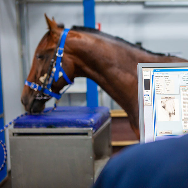
At the Ballarat Veterinary Practice, we have an in-house digital radiography system which allows us to take comprehensive sets of x-rays in a minimal amount of time and produce images which are of the highest quality available in veterinary medicine. Our radiography suite includes a dedicated imaging room with non-slip flooring, air conditioning and an adjacent processing/editing area.
In the field, our ambulatory vets have state-of-the-art portable digital radiography capability which allows them to capture high quality images on your property without then need for you to transport your horse to the hospital.
For our racing clients we also offer full yearling sales radiographic support. For the vendor we can provide a full set of Repository x-rays of your yearlings and submit them online to your chosen sales company. For the purchaser, we offer a complete range of sales vetting services at all the major yearling sales in Australia and New Zealand.
Ultrasonography
Ultrasound imaging in veterinary medicine has provided practitioners with a unique diagnostic tool for diseases of soft tissue. It is a safe, non-invasive and often, portable imaging modality that allows visualisation of any soft tissue including, but not restricted to the tendons, larynx, lungs, heart, gastrointestinal tract, abdominal organs and the reproductive tract.
Ballarat Veterinary Practice boasts a portable reproductive ultrasound machine which can be used at our Clinic or on your property if you have a suitable crush available for rectal reproductive examination and pregnancy diagnosis.
The clinic is also equipped with a state of the art Ultrasound machine that is used exclusively in house. This machine also has Doppler flow suitable for more advanced cardiovascular assessment and ultrasound guided injections.
Scintigraphy
Nuclear scintigraphy (bone scan) is a diagnostic imaging technique which uses a radioactive isotope to detect areas where active bone modelling is taking place within the horse’s skeleton. When bone is damaged, the repair process results in increased bone turnover. The cells responsible for this increased bone activity are highlighted by the radioactive isotope. These areas appear as ‘hot spots’ on the images created when the Gamma camera collects the radioactive emissions.

Scintigraphy is therefore extremely helpful for detecting painful sources of lameness in horses undergoing modelling and is very accurate for showing “bone stress reactions or fractures”. Because the whole skeleton can be imaged, normally difficult areas to examine such as the spine, pelvis and hindquarters are easily viewed.
Specific indications include the diagnosis of lameness where the site of pain is known but radiographs are inconclusive. Scintigraphy is also an invaluable tool in horses where there are multiple sites of pain. Scintigraphy has become an integral part of lameness diagnosis at the Ballarat Veterinary Practice Equine Clinic.
The procedure:
Horses are usually admitted on the day prior to the planned scintigraphic examination at which stage a lameness evaluation is conducted. On the day of the scan, the horse will be injected with the isotope, technetium 99. It takes approximately 2 hours to achieve good distribution and attachment of the isotope on to bone tissue. Once the technetium has been absorbed by the bone the scan is performed. The gamma rays are collected by the gamma camera over a period of time, (generally 90 seconds per image). The image production requires that there is minimal movement of the body part being scanned, or the images produced will be ‘blurred’.
In order to produce high quality images all horses are sedated prior to scanning. The gamma camera is connected to a computer that uses the information collected to build up 2D pictures of the horse’s skeleton. These pictures are then analysed by the veterinarian.
Depending on the area to be scanned, the examination generally lasts between 1 and 3 hours. Due to radiation safety horses are kept in isolation for 24 hours after the injection of the isotope until the radioactive isotope is all but eliminated from the horse’s body.
By combining the scanning results with other imaging modalities such as digital radiography, ultrasonography and using diagnostic analgesia (nerve blocks), a more precise diagnosis is possible. This enables a more accurate estimate of prognosis, which in many cases is critical when dealing with equine athletes.
MRI
Magnetic resonance imaging (MRI) is a medical imaging technique that uses a magnetic field and computer-generated radio waves to create detailed images of the organs and tissues in the body. It is the gold standard imaging technique for the assessment of soft tissues.
Diagnostic in over 90% of cases, MRI can accurately identify the specific cause of lameness and the degree of pathology which leads to a full understanding of the injury. This means an optimal treatment plan can then be initiated immediately with confidence.
The advantages of MRI include:
- High quality images of bone and soft tissue of the lower limb
- Ideal imaging modality for the accurate diagnosis of conditions involving the feet
- Early accurate diagnosis
- Eliminates the risk of further damage
- Crucial information regarding diagnosis, management and long-term prognosis
- Allows accurate treatment plan to be formulated
MRI at the Ballarat Equine Clinic is able to be performed under carefully monitored sedation which allows the horse to remain standing throughout. This eliminates the risks associated with general anaesthesia and anaesthetic recovery from lateral recumbency. An added advantage of standing MRI is that patients can be admitted on a day procedure basis.
With a large equine community in the Ballarat area, BVP has recognised a high demand for accurate lameness diagnostic technology. This is why we are proud to offer diagnostic imaging of the foot and leg using the Hallmarq Standing Equine MRI to give racing, performance and leisure horses an early, safe and accurate diagnosis. The Hallmarq MRI scanner is located in a purpose designed building at the clinic and a team of vets has been trained to use the scanner and interpret the resulting images to the highest standard.
The Ballarat Equine Clinic is proud to be one of the few privately owned practices in the country to offer the benefits of standing MRI.
The Hallmarq Standing Equine MRI uses a strong magnetic field to produce images of soft tissues and bone. The procedure is non-invasive and does not require general anaesthesia. Sedation is used to prevent the horse from moving throughout the procedure. In most cases we will remove the front or back shoes depending on the region of interest to prevent interference with the magnetic field. The scan itself takes around 1-2 hours and you may be able to pick up your horse later the same day depending on when the scan takes place.
However, each scan produces up to 500 separate images, so interpretation takes time. The images are sent to a specialist for interpretation and the diagnosis may take a few days.
MRI is used in conjunction with a thorough clinical examination protocol and is considered particularly useful where:
- Chronic lameness has been localised to the foot or lower limb by nerve blocks
- Radiographs are negative or equivocal
- Nuclear scintigraphy (bone scan) is being considered or is negative
- To more accurately define suspicious findings on radiography or scintigraphy eg pre fracture pathology
- Access to the area of interest by ultrasound is difficult or impossible due to hoof capsule
- There is a penetrating injury needing urgent attention
- Treatment and healing need to be monitored before returning to work
It is important to keep in mind that Standing MRI does not replace the skill and experience of an equine vet. The initial clinical examination is still essential and preliminary diagnostic imaging (ultrasound, x-ray, bone scan) may be recommended.
That said, MRI has been a powerful addition to our diagnostic imaging capabilities.
If you would like more information about MRI please call (03) 5334 6756.
Gastroscopy
Gastroscopy involves the examination of the equine oesophagus, stomach and proximal duodenum using a 3m long video endoscope and is the gold standard in the diagnosis of gastric ulcers in the horse.
Feed is withheld for 12-18 hours prior to gastroscopy which is typically performed under sedation. The mucosa of the oesophagus, stomach and duodenum is viewed and assessed.
Then a treatment regimen can be developed to resolve any mucosal lesions as quickly as possible. Repeat gastroscopes allow the response to treatment to be assessed.
URT Endoscopy
Horses are prone to disorders of the respiratory tract.
Endoscopic examination of the upper respiratory tract (URT) of the horse covers the nasal passages, pharynx, larynx and trachea. Video endoscopy enables the veterinarian to thoroughly examine the URT for any abnormalities.
Coughing, nasal discharge, epistaxis (nose bleeds) abnormal respiratory tract noise, facial swellings, respiratory distress and poor performance all warrant an endoscopic examination of the URT in order to make an accurate diagnosis. Typically, such cases are scoped at rest, whilst standing in a crush. During the endoscopic examination fluid samples can be taken from the respiratory tract and sent to an external laboratory for analysis.
However, a number of upper respiratory tract problems associated with abnormal anatomical function are induced by strenuous high intensity exercise and so may not be evident during a resting endoscopic examination.
Dynamic upper respiratory tract videoendoscopy (DVE) is now the gold standard in diagnosing abnormal function of the URT in the equine athlete. These scopes are performed during exercise. It allows the veterinarian to assess the horse’s URT and its response to the demands placed upon it during performance. DVE allows a more thorough evaluation of URT function and the extent to which it is affecting the horse’s ability to breath and perform.
Conditions such as low grade recurrent laryngeal nerve neuropathy (RLN), intermittent displacement of the soft palate, pharyngeal collapse and the functionality of other soft tissues can be easily diagnosed and documented in this way.
There are two types of dynamic upper respiratory tract video endoscopy, ‘Treadmill DVE’ and ‘Over Ground DVE’
Treadmill studies are routinely performed at BVP and will show most abnormalities., particularly soft palate dysfunction.
Over Ground DVE allows the horse to be examined in a way that more closely mimics race day conditions. The horse can be worked over varying distances, and with other horses which may provide an advantage over treadmill studies. Overground DVE is also an excellent tool for the valuation of sport horse’s where head and neck position and rider interaction have been shown to influence upper airway function. Watch this space for the introduction of overground DVE at BVP.
The Ballarat Veterinary Practice Equine Clinic has over 20 years’ experience performing and interpreting dynamic upper respiratory tract video endoscopy and the surgical expertise to address any abnormalities diagnosed.
