Equine Surgery
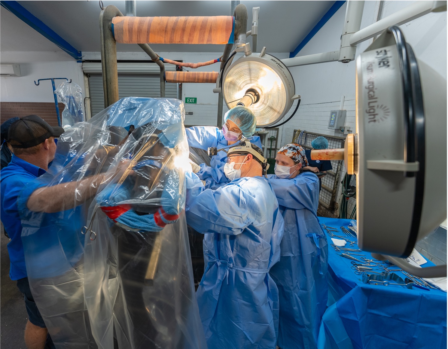
The Ballarat Equine Clinic offers a full range of primary care, referral and emergency surgical services.
- Comprehensive admission process and pre-operative evaluation with an individual treatment surgical and anaesthetic recovery plan for each surgical patient.
- Warm supportive waterbed and state of the art surgical table
- Dedicated veterinary anaesthetic monitoring
- Board certified surgical team with over 30 year’s experience
- Qualified and highly skilled surgical nursing team
- Video monitored post-operative recovery with option for sling recovery
- Facilities, bed, BP monitoring, anaesthetic equipment
Orthopaedic Surgery
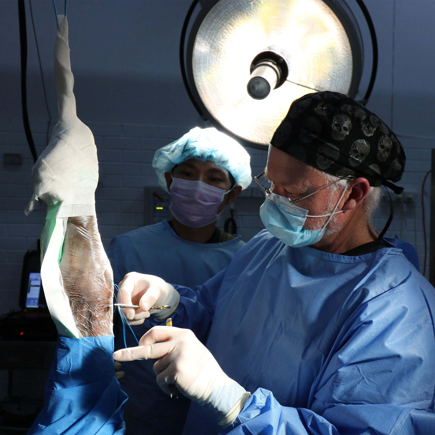
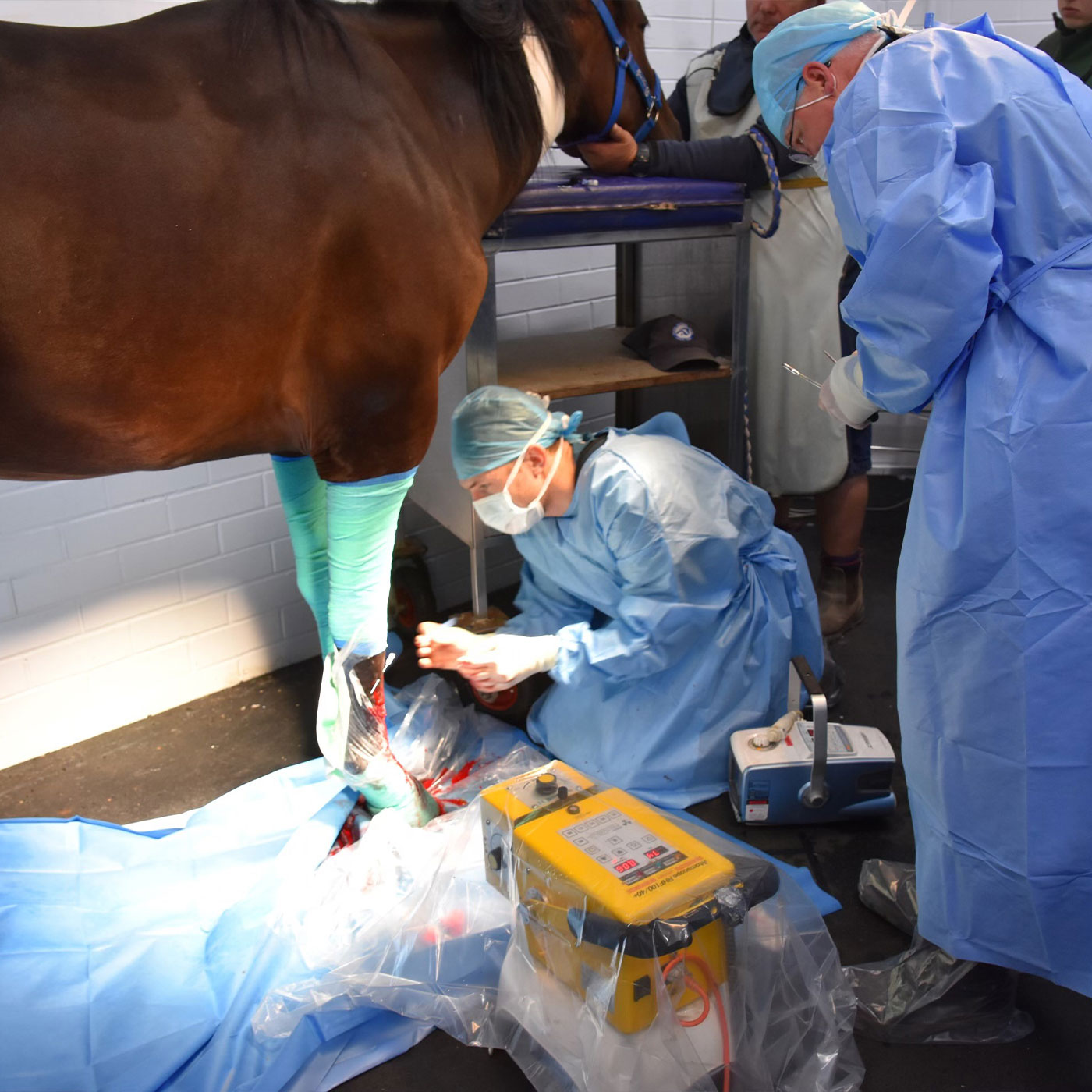
If possible surgery will be performed standing under carefully monitored sedation in order to avoid the risks associated with a general anaesthesia.
Orthopaedic Surgery / Arthroscopy
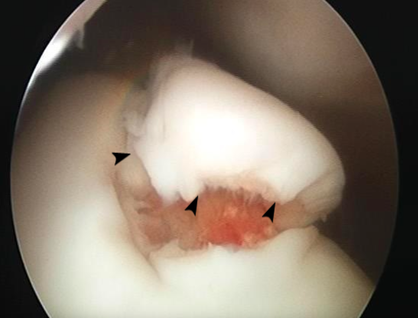
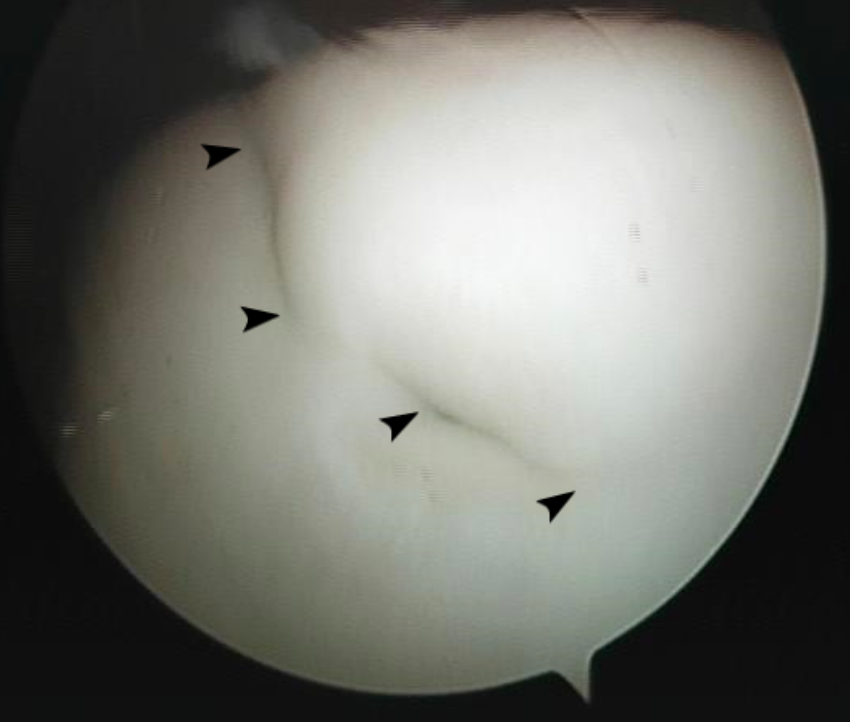
Arthroscopy is a surgical technique used by equine surgeons to explore, evaluate and treat joint disorders.
A narrow, rigid fibreoptic instrument called an endoscope is placed through a small incision (~1cm) into the joint space. An image is then transmitted to a camera for viewing on a monitor. In this way the surgeon is able to visualise and examine the various structures inside the joint in a minimally invasive way.
Specially designed surgical instruments are then passed through separate small incisions to assist in the repair of any joint pathology.
Commonly, arthroscopy is used to treat chip fractures, OCD lesions, subchondral bone cysts, open joint wounds, septic joints and assist in the repair of articular fractures (fractures involving the joint). Additionally, arthroscopy can be used to assess and treat diseased or damaged soft tissue structures within the joint.
Similar techniques are also used to evaluate and treat disease within tendon sheaths (known as tenoscopy) and bursae (bursoscopy).
Orthopaedic Surgery / Angular Limb Deformities (ALD)
Conformational abnormalities of the equine limb that are evident when assessed from the front (frontal plane) are known as an angular limb deformity (ALD).
Angular limb deformities are a common occurrence in foals. Some foals are born with an obvious ALD (perinatal deformities), others will acquire them. However, if intervention is provided in a timely fashion, it is possible in many cases to improve the foal’s conformation and thus help protect its future athletic potential.
From birth to weaning, your foal’s carpal, fetlock and foot conformation may change substantially.
For this reason, it is important to have your foal’s conformation assessed by your veterinarian at birth and reassessed every 2-4 weeks if there are any issues. Your veterinarian will determine how best to manage any ALD. Stall rest, controlled exercise, appropriate foot care and/or other non-invasive techniques may be all that is required. However, in some cases surgical intervention is necessary.
It is important to note that there is only a short window of opportunity in which it is possible to effectively make such surgical corrections. Beyond a certain age, such procedures will not result in the degree of correction required to promote long-term soundness. If surgery is indicated, it needs to be performed before the relevant growth plate/s have closed.
Surgical procedures to correct angular limb deformities need to be carefully planned and executed for the best anatomical and cosmetic result and ultimately for the patient’s long-term soundness.
Ballarat Veterinary Practice offers periosteal stripping as well as transphyseal bridging to correct the more severe angular limb deformities.
Orthopaedic Surgery / Fracture Repair
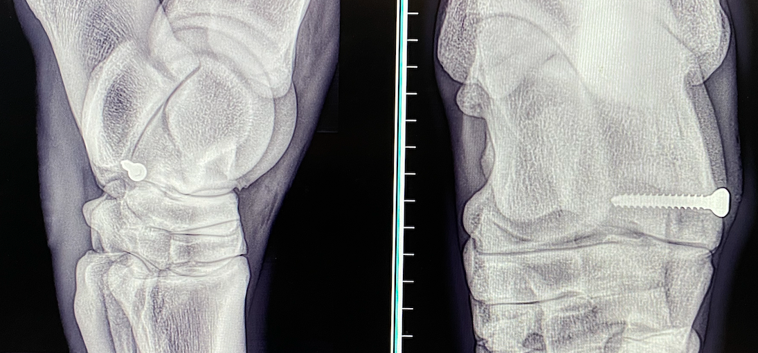
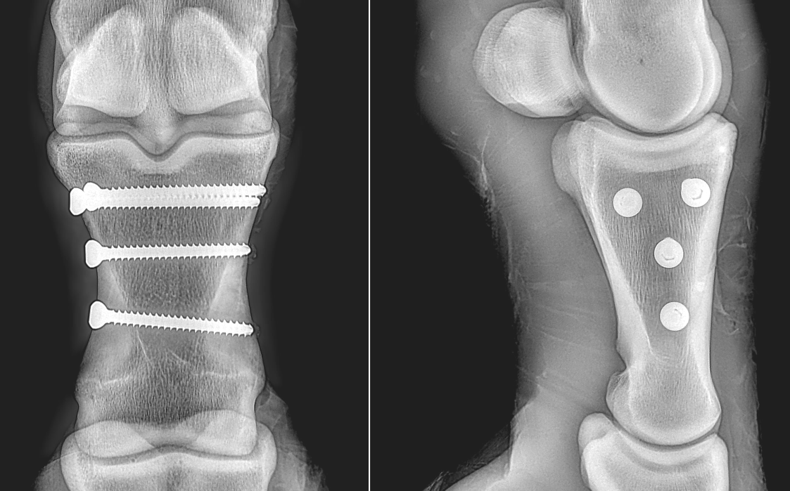
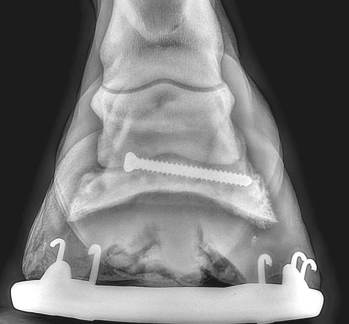
Our surgeons are highly trained in techniques for the application of bone screws and plates for the repair of equine fractures. Following assessment with radiographs (x-rays) a surgical plan is made. For simple non-displaced fractures, bone screws can be used and it is not uncommon for such horses to return to an athletic career. The use of bone plates can also be successful.
Orthopaedic Surgery / Kissing Spine Surgery
In normal horses there is a space present between the bones of the thoracic region of the vertebral column, i.e. beneath the saddle. When there is no space or the bones are too close together this results in the condition known as ‘kissing spine.’ In many cases this condition is painful and results in poor performance, lameness and or behavioural issues.
Whilst a number of patients will respond well to conservative therapy (anti-inflammatories, rest and physical therapy) surgical intervention in refractory cases may be necessary to improve their quality of life. Many horses that have had kissing spine surgery have subsequently returned to the competition arena and gone on to have successful careers.
Soft Tissue Surgery / Colic Surgery
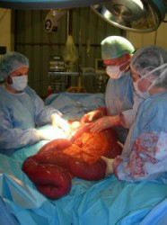
Ultimately, a horse that continues to show signs of abdominal pain in the face of medical treatment and pain relief requires surgical exploration of the abdomen (exploratory laparotomy). An exploratory laparotomy allows the surgeon to visualise and assess the entire intestinal tract and abdominal organs and diagnose and where possible treat the cause of the horse’s pain. Fortunately less than 10% of horses presented with colic will require surgical intervention.
From simple impactions and intestinal displacement, to strangulating lipomas and severely compromised intestinal blood flow, there are many conditions that require surgical correction (see below).
Once the procedure required to correct any abnormalities is identified the surgeon will be able to give you a definitive diagnosis and an informed assessment of the likelihood of a successful surgical outcome. In this way, you can make the best decision for your horse.
It is important to note that in many cases, horses respond well to surgery and go on to return to full athletic careers upon recovery.
If your horse is suffering from unrelenting pain and surgery is not possible, then with the utmost concern for your horse, euthanasia will be recommended.
Soft Tissue Surgery / Laparoscopy
Minimally invasive surgical techniques performed via laparoscopy, lead to decreased soft tissue trauma, reduced postoperative pain and complications. Laparoscopic surgery typically leads to improved post-operative outcomes and shorter recovery times which may allow the patient to return to normal function sooner.
Where once many abdominal surgical procedures required a large midline incision to enable the surgeon to access the organ or pathology of interest, laparoscopy allows visualisation of the internal equine abdomen via an endoscope which is passed into the abdomen through small incisions known as portals. Often laparoscopy offers better visualisation of the equine abdomen.
Laparoscopy is typically performed as a standing (hyperlink please) procedure under carefully monitored sedation. The abdomen is insufflated (distended) with gas, usually carbon dioxide, and instruments are inserted through portals in the flank of the horse. A video camera attached to the viewing laparoscope allows the surgeon to visualise the internal abdomen on a monitor.
Laparoscopy can be used both for diagnostic purposes and surgical procedures. Diagnostically, laparoscopy can be used to investigate chronic weight loss, chronic colic, intra-abdominal haemorrhage, neoplasia and peritonitis.
Surgical laparoscopy is ideal for cryptorchid castration (removal of abdominal testis), ovariectomy, ruptured bladder repair, Urinary calculi removal, hernia repair, ovarian granulosa theca cell tumour removal and the breakdown of abdominal adhesions.
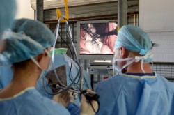
Standing laparoscopic procedure
Upper Respiratory Tract Surgery
The Ballarat Veterinary Practice specialises in Upper Respiratory Tract Surgery. Our highly experienced surgeons frequently perform surgeries for:
- “Roaring” or laryngeal paralysis (recurrent laryngeal neuropathy) – laser, tie-back, nerve pedicle graft surgery
- Arytenoid Chondritis – laser and arytenoidectomy
- Epiglottic Entrapment – splitting surgery, laser resection, laryngotomy
- Soft Palate Displacement – palate cautery, tie-forward surgery
- Dynamic airways collapse – laser surgery
We can frequently perform these procedures in the sedated standing horse which avoids the risks associated with general anaesthesia and allows a quicker immediate post operative recovery.
